Diagram Of Shoulder Bones / Shoulder Joint Anatomy Pictures And Information - As a ball and socket synovial.. Each vertebra has a hole in it. Consisting of the clavicle (collar bone) and scapula (shoulder blade), the pectoral girdle forms the attachment point between the arm and the chest. The shoulder is not a single joint, but a complex arrangement of bones, ligaments, muscles, and tendons that is better called the shoulder girdle. When this type of cartilage starts. Placebo injection for adhesive capsulitis of shoulder.
The shoulder bones, rib bones and hip bones ,are all joined to the backbone. Growth occurs when cartilage cells divide and increase in number in these growth plates. As a ball and socket synovial. Three bones in the fetus develop into the humerus bone in adults. Related posts of diagram of shoulder muscles and tendons.
Illustration Of The Bony Anatomy Of The Shoulder Joint Complex Download Scientific Diagram from www.researchgate.net The shoulder is not a single joint, but a complex arrangement of bones, ligaments, muscles, and tendons that is better called the shoulder girdle. When this type of cartilage starts. Shoulder problems including pain, are one of the. A normal, complete, human skeleton includes two shoulder blades, which the scapula is a flat bone so it is conveniently described by two diagrams, one to label the features of the posterior surface of the scapula and the. In human anatomy, the shoulder comprises the part of the body where the arm attaches to the torso. Human science human body arm anchor chart bone shoulder health skeleton diagram. These are the supraspinatus, infraspinatus, teres. Lower jaw (mandible) collar bone.
The human shoulder is made up of three bones:
Each vertebra has a hole in it. Growth occurs when cartilage cells divide and increase in number in these growth plates. The shoulder is not a single joint, but a complex arrangement of bones, ligaments, muscles, and tendons that is better called the shoulder girdle. Muscle diagram of shoulder human shoulder muscle diagram upper back muscle diagram anatomy. Posterior to the clavicle is the scapula, a flat, triangular bone located lateral to the thoracic spine in the dorsal region of the body. The first type is the white cartilage on the ends of the bones (called articular cartilage) which allows the bones to glide and move on each other. Human science human body arm anchor chart bone shoulder health skeleton diagram. Shoulder problems including pain, are one of the more common reasons for physician visits for musculoskeletal symptoms. Back of skull (occipital bone). The transverse humeral ligament is not shown on this diagram. Click now and learn everything about its anatomy and function at kenhub! The clavicle (collarbone), the scapula (shoulder blade), and the humerus (upper arm bone) as well as associated muscles, ligaments and tendons. The shoulder joint is formed where the humerus (upper arm bone) fits into the scapula.
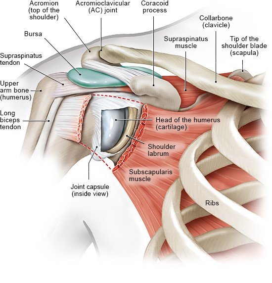
As a ball and socket synovial. Cheek bone (zygoma) upper jaw (maxilla). This image shows the shoulder joint displaying the bones and ligaments that form the joint and supports it (from anterior view) showing: Simple easy notes for quick revision for exams. A long bone, such as your femur (thigh bone), grows in length at either end in regions called growth plates.
How Does The Shoulder Work Informedhealth Org from www.informedhealth.org A regional atlas of the human body is 7 draw labelled diagram showing the relations of shoulder joint. The shoulder joint (glenohumeral joint) is a ball and socket joint between the scapula and the humerus. Growth occurs when cartilage cells divide and increase in number in these growth plates. There are two kinds of cartilage in the joint. Click now and learn everything about its anatomy and function at kenhub! Placebo injection for adhesive capsulitis of shoulder. Each vertebra has a hole in it. The transverse humeral ligament is not shown on this diagram.
When this type of cartilage starts.
Shoulder flexion is movement of the shoulder in a forward motion. The scapula is the flat triangular bone which provides an attachment for muscles for the shoulder, back and neck. Shoulder diagram illustrations & vectors. The main functions of shoulder bone are : An example of shoulder flexion can be seen when reaching forward to grasp an object. Back of skull (occipital bone). Shoulder bones and ligaments anatomy. Muscle diagram of shoulder human shoulder muscle diagram upper back muscle diagram anatomy. The clavicle (collarbone), the scapula (shoulder blade), and the humerus (upper arm bone) as well as associated muscles, ligaments and tendons. The first type is the white cartilage on the ends of the bones (called articular cartilage) which allows the bones to glide and move on each other. The shoulder is the most movable joint in the body. The transverse humeral ligament is not shown on this diagram. The scapula is a large, flat triangular bone with three processes called the acromion, spine and coracoid process.
A normal, complete, human skeleton includes two shoulder blades, which the scapula is a flat bone so it is conveniently described by two diagrams, one to label the features of the posterior surface of the scapula and the. The shoulder is not a single joint, but a complex arrangement of bones, ligaments, muscles, and tendons that is better called the shoulder girdle. In human anatomy, the shoulder comprises the part of the body where the arm attaches to the torso. The shoulder joint is formed where the humerus (upper arm bone) fits into the scapula. Shoulder joint is the most mobile joint of the human body.
Shoulder Anatomy from fpnotebook.com Printable shoulder muscles diagrams to help you study the muscles structure in human's shoulder.we have five muscle diagrams of the shoulder. This image shows the shoulder joint displaying the bones and ligaments that form the joint and supports it (from anterior view) showing: Due to this, there is a hollow centre inside the backbone. When this type of cartilage starts. There are two kinds of cartilage in the joint. (1) collar bone on the two sides of the next keep our shoulders apart. Each vertebra has a hole in it. These are the supraspinatus, infraspinatus, teres.
A regional atlas of the human body is 7 draw labelled diagram showing the relations of shoulder joint.
A long bone, such as your femur (thigh bone), grows in length at either end in regions called growth plates. Placebo injection for adhesive capsulitis of shoulder. Hand drawn realistic human bones. Following inferior dislocation of shoulder joint, the rounded contour of shoulder is lost and there is weakness of abduction of armbecause the. In human anatomy, the shoulder comprises the part of the body where the arm attaches to the torso. Lower jaw (mandible) collar bone. The shoulder joint (glenohumeral joint) is a ball and socket joint between the scapula and the humerus. Download 708 shoulder diagram stock illustrations, vectors & clipart for free or amazingly low rates! Click now and learn everything about its anatomy and function at kenhub! Scapula (= 'shoulder blade' or 'shoulder bone') is a bone of the human body. Related posts of diagram of shoulder muscles and tendons. Due to this, there is a hollow centre inside the backbone. Shoulder problems including pain, are one of the.
The shoulder is the most movable joint in the body diagram of shoulder. This page is about shoulder bone anatomy diagram,contains anatomy of the shoulder central coast orthopedic medical group,anatomy of the shoulder part 3 (muscular structures),shoulder replacement,guide to shoulder anatomy and more.


0 Komentar